Next topic Previous topic Table of Contents
A nerve is a bundle of nerve fibers. The nerve impulse evoked by stimulation of a nerve is a collective event recorded as it engages as many fibers as may be recruited by the intensity of the stimulus. A reasonably homogeneous subpopulation of those fibers will yield an external record comparable in form to the external record from a single axon.
Topic 13: The subject: the sciatic nerve isolated from a frog leg
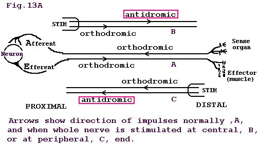
The sciatic nerve is a large bundle of many nerve fibers. The fibers come out between vertebrae at the caudal end of the vertebral column. The nerve of the frog is dissected from its origin at the spinal cord as 3-4 bundles of the sciatic plexus, all the way to the gastrocnemius muscle. There the nerve divides into two branches, one to each head of the muscle. As we proceed distally along the nerve, from the plexus, we find fibers leaving the main trunk and entering muscles and skin. Therefore, having fewer fibers, the distal end of the nerve is of smaller diameter than the proximal end at the plexus. The fibers that normally carry impulses from the central nervous system (CNS) outward to the periphery to muscles and skin are efferent or motor. Impulses normally move toward the CNS along afferent or sensory fibers from peripherally located sensory receptors in muscle and skin.
Afferent fibers are dendrites, and efferent fibers are axons, but any peripheral nerve fiber of unspecified nature is commonly called an axon. If the nerve is stimulated at the proximal end, impulses travel in the normal direction (orthodromically) in the efferent fibers but antidromically in the afferent fibers. That is, impulse propagation can be induced to proceed equally well in either direction along a nerve fiber. Advantage is taken of this fact in the sciatic nerve taken out of a frog leg.
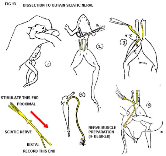
The sciatic nerve is dissected free from the leg of a frog and is immersed in a bath of physiological solution. The bath is provided with electrodes for initiating and recording action potentials. The dissection procedure is illustrated in Fig. 13.
Figure 13. Preparation of frog nerve.
Next topic Previous topic Table of Contents
Customarily, a physiological (Ringer’s) solution to sustain isolated tissues is made by mixing suitable amounts of the required salts to approximate blood. A mixture of one part seawater to three parts distilled water is less trouble to prepare and serves very well. Except for the Mg++, the ratios of the inorganic salts are nearly the same in sea water as in blood. The high Mg++ does not appear to affect impulse propagation significantly. If need be, the Mg++ can be counteracted by the addition of extra Ca++.