Upper tracings: membrane potential outside of the fiber is arbitrarily held constant at zero. Inside resting potential is negative and moves in positive direction during AP. This is probably the most popular convention.
Next topic Previous topic Table of Contents
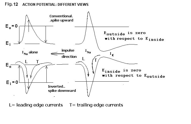
Representing time increasing from left to right, in Fig. 12, we show the membrane potential inside of the membrane with respect to that outside considered zero. It can just as well be shown as the change in outside potential compared to that inside, i.e., inverted. In either case, the impulse direction of propagation is from right to left in this figure. The front, or leading edge, of the action potential, is at the left, the trailing edge at the right.
Figure 12. The action potential: different views.
Upper tracings: membrane potential outside of the fiber is arbitrarily held constant at zero. Inside resting potential is negative and moves in positive direction during AP. This is probably the most popular convention.
Lower tracings: inside of fiber is arbitrarily held constant at zero. Outside then goes negative during AP. This assumption is just as "correct" as the above. Whichever convention is used, try to be consistent in order to avoid confusion.
At right: AP has no after-hyperpolarization because |EM|=|EK|
At left: AP has after-hyperpolarization because |EM|>|EK|
The action potential is a traveling wave of depolarization moving along the nerve fiber. As the zone of maximum depolarization moves along, it serves as a sink for depolarizing current ahead, at the leading edge.
The sink also draws current at the trailing edge, where repolarization is occurring. Thus the longitudinal currents reflect the longitudinal distribution of the membrane potential gradient (see Figs. 10, 11).
The lesson to this point has been an introduction to the transmembrane action potential of the single nerve fiber. The collective activity of many fibers in a nerve can be monitored by the currents that flow externally. Examples obtained from isolated sciatic nerve of the frog Rana pipiens will be presented.
Next topic Previous topic Table of Contents