Fig. 32A. S2-EPP sums with decaying
S1-EPP to yield AP.
S1-EPP to yield AP.
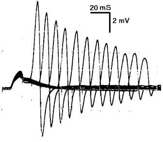
Fig. 32B. Decay of EPP facilitation.
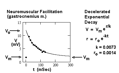
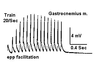
Fig. 32C. EPP facilitates nonpropagated AP.
Next topic Previous Topic Table of Contents
|
Fig. 32A. S2-EPP sums with decaying
S1-EPP to yield AP. |
|||
|
|
Same as 32A, except responses to increasing C-T intervals are shown. The amplitude of the second response (AP) decreases monotonically with interval since the first stimulus. | ||
|
Fig. 32B. Decay of EPP facilitation.
|
|||
|
|
At long C-T interval, the EPP evoked by S1 has decayed to a negligible level, and the test response from S2 approaches the amplitude of a single response. The data are modeled by a decelerated exponential decay function. The decay of AP peak reflects time course of the single EPP. | ||
|
Fig. 32C. EPP facilitates nonpropagated AP.
|
|||
|
|
In this example, a spike indicative of a muscle AP is generated by each stimulus during a train of stimuli to the nerve. There is no reversal of current direction, so the AP does not appear to be propagated. |
Next topic Previous Topic Table of Contents